


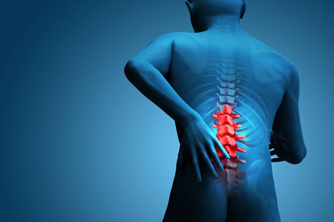
Osteochondrosis in the lumbar region occurs due to deformation and destruction of the cartilaginous tissue of the intervertebral discs.Without cartilaginous material, the vertebrae lose their elasticity and shift, causing pain.According to the WHO, the disease mainly affects people over 30 years of age.Women get sick three times more often, which is also associated with age-related changes and a decrease in calcium levels in the body.
Osteochondrosis of the lower back.
Of all diseases of the peripheral nervous system, osteochondrosis of the spine accounts for 80%, of which 60-80% affects the lumbar region, which consists of 5 vertebrae.In 40% of people, the first signs of the disease appear between the ages of 30 and 35.In old age, spinal osteochondrosis is diagnosed in 90% of people.7 out of 10 people experience painful sensations;The exacerbation can last from 6 to 16 months.
Development stages
- 1st – the appearance of degenerative disorders, which affect nerve endings and alter blood supply.The inflammatory process causes pain that radiates to the legs.The patient experiences occasional tingling in the back and buttocks.It appears after intense physical effort.
- 2nd – The outer layer of the intervertebral disc (annulus fibrosus) is destroyed.The distance between the vertebrae is reduced, the nerve endings are compressed, which causes intense, sharp pain.The symptoms are especially pronounced when walking.
- 3rd – due to excessive mobility of the vertebrae, muscle fibers and nerve endings are compressed.Spasms, numbness, burning, hernias occur - displacement of the intervertebral disc towards the spinal cord.
- 4th – growth of pathological exostoses in the bone structure.The spine literally "turns to stone."Progressive osteoarthritis and lack of treatment immobilize the patient.
Reasons for development
The disease develops slowly, under the confluence of a number of circumstances, that is, the development of the disease depends on the influence of not one, but several factors:
- Sedentary lifestyle (physical inactivity), sedentary work, low level of physical activity.At first glance they are not dangerous.However, with prolonged exposure, muscle tone, the so-called "muscle corset", weakens.Supports the spine and internal organs.Therefore, physical inactivity causes an alteration in the functioning of several systems: blood circulation, breathing and digestion.
- Overweight.With a BMI (Quetelet body mass index) greater than 25 kg/m2, excess weight creates additional stress on the body: it puts pressure on the spine, overloads the joints and impairs blood supply.
- Endocrine pathological processes.Hormonal imbalance, disturbances in the functioning of the adrenal glands and thyroid "slow down" the body's metabolism.Frequent hormonal changes "wash out" calcium from bone tissue, leaving it weak and brittle.
- Unbalanced diet.Popular and affordable fast food fully satisfies the energy needs of the body, while from the point of view of saturating the body with the necessary vitamins and microelements, it is absolutely useless.
- Pathologies and genetic predisposition to diseases of the musculoskeletal system.
- Age-related degenerative changes.Under the influence of time, natural wear of the support system occurs.
In men whose profession involves significant physical activity, lumbar osteochondrosis occurs many times more often.
Symptoms of lumbar osteochondrosis.
Pain is the main marker of the onset of the disease and its location.Depending on the nature of the occurrence, frequency and duration, a preliminary diagnosis can be made.Most often, patients with lumbar osteochondrosis experience pain that radiates to the leg, groin and tailbone.In a horizontal position, the discomfort disappears.
Spinal nerves extend from the trunk of the spine to different parts of the body.Each nerve is responsible for a specific area.This is called "segmental innervation."This is why the affected nerve “responds” in a specific area: this is how “radicular syndrome” manifests itself.
The spinal nerve map allows you to identify pain in a specific area of the spine: C1-C8 - 8 cervical, T1-T12 - 12 thoracic, L1-L5 - 5 lumbar, S1-S5 - 5 sacral, C0 - 2 coccygeal.Signs of osteochondrosis depending on segmental innervation:
- L3-L4 – weak knee reflex, partial numbness, episodic loss of sensation are observed along the anterior surface of the thigh;
- L4-L5 – muscle weakness, the patient cannot fix the body while standing on the heels, pain and impaired sensitivity extend from the lumbar area to the buttocks, as well as along the entire lateral surface of the thigh (from top to bottom).
- L5–S1: Calf muscle tremors, inability to stand on tiptoe, pain in the back of the thigh.
In rare cases, the root arteries also participate in the destructive process, causing a spinal stroke - a dangerous pathology in which the patient experiences paresis and paralysis, as well as dysfunction of the pelvic organs.
Pain reflex symptoms.
- Lumbago or lumbago (in this case not sciatica!): acute pain.Any uncomfortable movement, even sneezing and coughing, causes a spasm.The patient spontaneously changes the position of his body, moving it to the side.An attempt to adopt a vertical position causes new lumbago.
- Lumbodynia is a dull, aching pain that occurs on one side.Periods of remission are followed by an exacerbation, which gradually turns into severe pain.
- Lumboischialgia: painful sensations appear in the leg.
Secondary signs of the disease are not so typical and can be easily confused with symptoms of another problem: poor sweating (hypohidrosis), pale skin in the affected area due to poor blood circulation, severe dryness and peeling.
Diagnosis
At the initial stage, a complete medical history of the patient is collected.The general picture must be complemented by the opinions of a surgeon, orthopedist, neurologist and rheumatologist.Specialists subject the patient to a visual examination to evaluate changes in posture, muscle tone, and skin sensitivity.
X-ray is performed for the primary diagnosis of lumbar osteochondrosis of the first degree;It is used to examine the condition of the intervertebral discs, pathologies of the spinal cord, and also to evaluate the general condition of the spine.However, this type of diagnosis does not provide complete information about the affected tissues.
Computed tomography (CT) is a highly sensitive test that uses a beam of ionizing rays.Computed tomography records the degree of disc deformation, inflammatory processes in the spinal cord membrane and nerve endings in a layer-by-layer image.
Magnetic resonance imaging (MRI) informatively shows the general condition of muscle mass, blood vessels and ligaments.Detects tumors and inflammatory processes.
Myelography is one of the ways to visually assess the condition of the spinal cord and nerve roots by injecting a contrast agent with a needle into the cavity between the pia mater and the arachnoid of the spinal cord, filled with cerebrospinal fluid.Like CT, MTP, and X-ray, the procedure is performed in real time.
Neurological examination of sensitivity and reflexes in pathologies is a complex of studies that includes a screening method for studying the symmetry of the limbs, testing sensitivity to vibrations and evaluating Babinsky, Chaddock and Oppenheiman reflexes.
Treatment of lumbar osteochondrosis.
The effectiveness of conservative treatment is achieved by a combination of drugs from various groups, massage and manual therapy.However, at stage 3, lumbar osteochondrosis can no longer be cured with conservative methods;Surgical intervention is required.One of the surgical methods for osteochondrosis is microdiscectomy.This is a neurosurgical operation to remove a hernia without damaging nerve structures.It is performed under general anesthesia.The patient can walk on the third day.
Drug therapy (for stages 1-2)
- Analgesics to relieve pain: local (ointments, gels), oral (tablets, suspensions), intramuscular (injectable solutions).
- Non-steroidal anti-inflammatory drugs to prevent the development of the inflammatory process.Reduces temperature, eliminates swelling.Most often gels and tablets are used, in more severe cases (inflammation of the sciatic nerve) - injections.Prima Course - 7-14 days.
- Muscle relaxants.It is prescribed in the acute period for muscle spasms, as well as during the period of remission to consolidate the effect.
- Chondroprotectors.Blocks destructive processes in intervertebral discs, promotes regeneration, improves metabolism in cartilage tissues.
- Vascular drugs.Helps normalize blood supply to the musculoskeletal system.
- Group B vitamin complexes. They improve nerve conduction.
- Diuretics.Reduces swelling.
As emergency help at the time of an exacerbation, powerful injections are used - paravertebral blockade, which causes a temporary loss of sensitivity.
It is also recommended to undergo manual therapy treatment twice a year, which relieves muscle tension and spasms, restores blood circulation, improves lymphatic circulation and increases the mobility of the musculoskeletal system.
Prevention
- Active lifestyle combined with moderate physical activity;
- Choose comfortable footwear or, if indicated, orthopedic;
- Good nutrition;
- Body weight control;
- Sleep only on an orthopedic mattress and pillow;
- Office furniture with flat backrest only;
- Rejection of heavy loads on the back and spine.When carrying heavy objects, the load should be distributed evenly between both hands.